BMI (Body Mass Index) was developed in the 1830s as a population-level statistical tool. It was never intended for individual clinical assessment. Yet it has become the dominant metric by which weight-related health risk is assessed. The problem is fundamental: BMI measures mass relative to height, with no information about body composition, fat distribution, or metabolic risk.
The Two Types of Body Fat
Not all fat is metabolically equivalent. Subcutaneous fat, the fat under the skin, is metabolically relatively inert. It contributes to body weight and body image, but its direct metabolic impact is limited. Visceral fat, the fat stored within the abdominal cavity surrounding the liver, pancreas, kidneys, and intestines, is an entirely different matter.
Visceral adipose tissue is metabolically active. It releases a cocktail of inflammatory cytokines and adipokines (including TNF-alpha, IL-6, and resistin) that directly drive insulin resistance, systemic inflammation, hypertension, dyslipidaemia, and non-alcoholic fatty liver disease. It is visceral fat, not subcutaneous fat or total body weight, that most strongly predicts cardiovascular and metabolic disease risk.
The phenomenon of ‘metabolically obese normal weight’ (MONW), sometimes called ‘skinny fat’, is well-documented. Individuals with a normal BMI but high visceral fat have metabolic risk profiles comparable to those who are clinically obese. Conversely, some individuals with elevated BMI but low visceral fat have near-normal metabolic risk. BMI tells you neither.
Why Standard Measurements Fail
Waist circumference and waist-to-hip ratio are improvements on BMI but remain crude. DEXA scanning quantifies body composition (fat mass vs lean mass) but cannot distinguish visceral from subcutaneous fat. The gold standard for visceral fat quantification is MRI, specifically cross-sectional abdominal imaging that measures the visceral adipose tissue area directly.
Multi-Organ MRI at Harley Weight Loss Clinic
Our multi-organ MRI protocol provides direct quantification of visceral fat compartments, alongside assessment of hepatic fat content, pancreatic health, and overall organ morphology. This transforms weight management from a subjective, scale-based exercise into a data-driven clinical programme with precise, measurable targets.
- Visceral fat area (VFA) quantified in cm², a direct metabolic risk marker
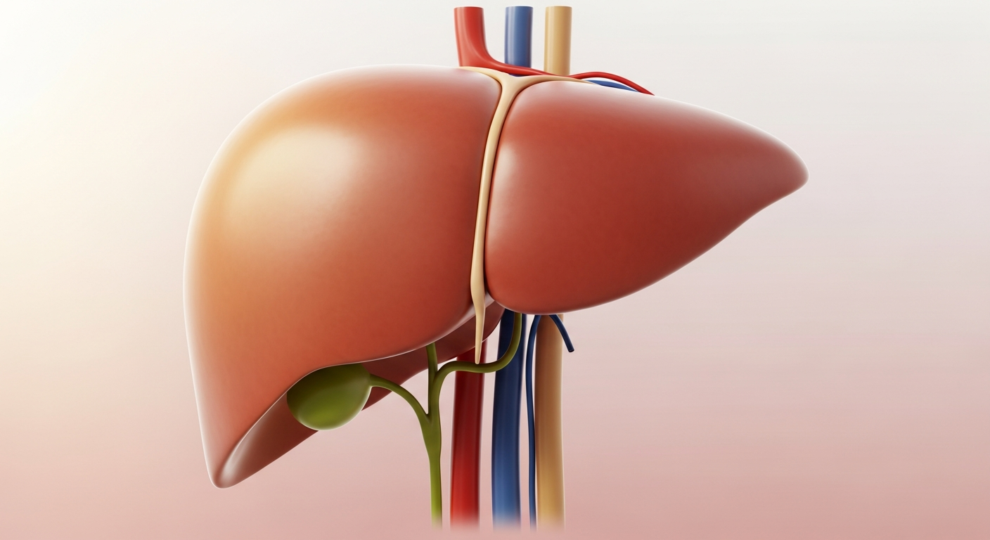
- Liver fat fraction, identifying NAFLD at the earliest stage
- Subcutaneous fat distribution, informing body composition targets
- Baseline imaging for longitudinal tracking of true clinical progress
- Incidental findings, occasionally identifying other clinically significant pathology
For patients who have struggled with conventional weight management approaches, or who want to understand their true metabolic risk rather than rely on an imprecise scale number, advanced imaging represents the clearest possible clinical picture. At our clinic, it is available as part of our comprehensive diagnostic pathway and can be arranged alongside an initial consultation with Dr Ajaz.
Written by Dr Saima Ajaz, MBBS, MRCGP
Lead Clinician, Medical Director, Harley Weight Loss Clinic